In addition to use of Optical Coherence Tomography (OCT) to image damage caused by glaucoma to the optic nerve and retina in our animal model, our program has established a number of interdisciplinary collaborations, including ongoing optical imaging projects that aim to gather qualitative and quantitative data relating to organization of the extracellular matrix, and development of aqueous outflow pathways, also involving the animal model at the center of our research. Dr. McLellan is currently the Director of an NIH –funded and supported shared resource that provides advanced imaging modalities to vision researchers on the UW- Madison campus.
Heidelberg Spectral Domain Optical Coherence Tomography
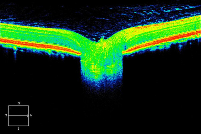
Heidelberg Spectralis HRA + OCT ® provides multi-modality imaging of the eye using a spectral-domain OCT integrated with confocal scanning laser ophthalmoscopy (cSLO). Multi-modality imaging offers insights into structure, function and metabolic activity of the retina. The Spectralis provides point-to-point correlation between fundus and OCT scans, producing an unmatched overall perspective of the retina and optic nerve head. This instrument is housed in Dr. McLellan’s Research space on campus at the University of Wisconsin-Madison and an additional instrument belonging to the Department of Ophthalmology and Visual Sciences (DOVS) is also available for use in animals by investigators within the Wisconsin Institutes for Medical Research (WIMR). This instrumentation is available to qualifying vision research investigators campus-wide including investigators from the UW McPherson Eye Research Institute (MERI), which supports the ongoing maintenance of this instrument. Preferential access is assured for NIH grantees named as users on the proposal that funded purchase of this instrumentAccess to this instrument represents an invaluable tool for NIH-funded investigators who study rodent and other animal models of ocular diseases ranging from retinopathies to glaucoma to corneal diseases.

The availability of the multi-modality imaging technologies incorporated in this instrument greatly facilitates the completion of existing projects but is also available to those wishing to generate preliminary data to support future proposals. Reliance on cross-sectional data is reduced, as the instrument enables longitudinal imaging studies –including fluorescein angiography and detection of fluorescent labels in retina tissues – to be carried out with precision in individual animal subjects non-invasively, in vivo. This improves quality and limits variability of data sets acquired, as well as reducing the number of animals required in our research programs. Technical support and training in data acquisition can be provided by the NEI-funded Vision Research Core. Requests to access the Heidelberg Spectralis HRA + OCT should be submitted to the NEI-funded Vision Research Core.